
Imaging Workflow
Provides a comprehensive, cloud-based infrastructure for managing patient data, imaging studies, and diagnostic workflows—securely and efficiently.
FDA 510(k) cleared · K250694
Canada's First FDA-Cleared AI for Intracranial Hemorrhage Triage. Our AI-powered detection delivers unparalleled accuracy and reliability with an average processing time of just 5.97 seconds.
Scaida Brain CT-ICH is a specialized AI module trained to detect various types of intracranial hemorrhages from non-contrast brain CT (NCCT) scans. It supports rapid, accurate triage and enhances clinician confidence when every second matters.
(Data collected from 6 institutions in the United States)
• Emergency response & trauma units
• Neurology & stroke centers
• Telestroke and teleradiology networks
One page on what BrainCT-ICH detects, how it notifies and what it takes to put it in front of your worklist.
Scaida BrainCT-ICH is AI software from mlHealth 360 for computer-assisted triage and notification of suspected intracranial hemorrhage (ICH) on non-contrast head CT. It flags suspected cases back to the radiologist’s worklist so they can be reviewed sooner.
Yes. Scaida BrainCT-ICH is FDA 510(k) cleared (K250694) for computer-assisted triage and notification of suspected intracranial hemorrhage on non-contrast head CT in adults aged 22 and above. It is not for diagnostic use and does not replace radiologist interpretation.
It flags suspected intracranial hemorrhage, which can be any of five subtypes: intraparenchymal (IPH), intraventricular (IVH), subarachnoid (SAH), subdural (SDH) and epidural (EDH) hemorrhage. It reports only whether ICH is suspected; it does not tell the user which subtype.
Eligible head CTs arrive from your PACS or RIS through standard DICOMweb, built-in checks route only eligible scans, and suspected ICH is flagged back to your worklist through a secure API. There is no new viewer and no second login, and it never alters images or removes cases from the queue.
In the retrospective study in its FDA 510(k) summary, sensitivity was 0.867 (95% CI 0.811–0.908), specificity was 0.887 (95% CI 0.812–0.934) and the AUC was 0.926. The study used 294 non-contrast head CT cases from three US institutions (two independent of the development data), acquired on GE Medical, Siemens and Toshiba scanners, with ground truth from three ABR-certified neuroradiologists.
Yes. It runs in the cloud or on-premises, in your environment or ours. On-premises deployment needs under 16 GB of GPU memory.
Teleradiology and reading groups, hospitals without overnight coverage and multi-site imaging groups, as well as emergency and trauma units, neurology and stroke centers, and telestroke networks.
Provides a comprehensive, cloud-based infrastructure for managing patient data, imaging studies, and diagnostic workflows—securely and efficiently.
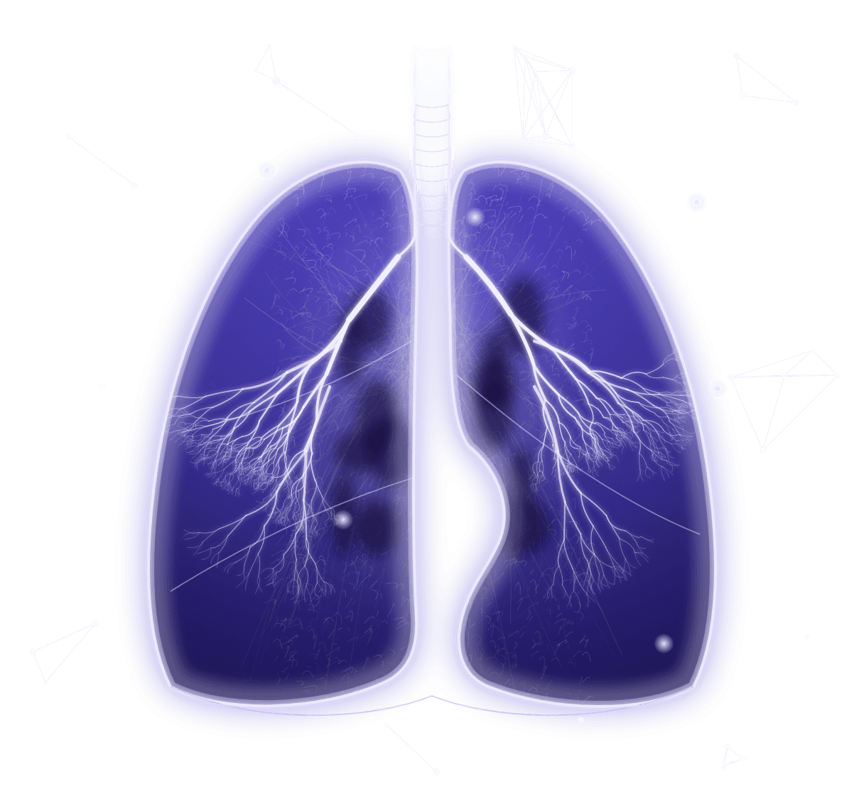
Reads a standard chest X-ray for 17 chest findings, flags them for a qualified reader and drafts the report.
Scaida
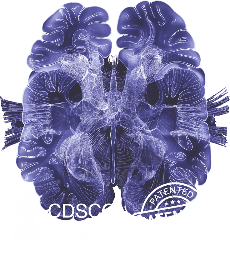
Provides precise brain segmentation and anomaly detection, assisting neuroradiologists in making life-saving decisions.

Messaging

Compatibility

Interoperability

security & scalability

Trained on real-world CT scans and expert-authored reports, this tool transforms AI insights into clinically accurate radiology reports
with our sales representative to discover
the perfect solution for your need